Orthodontics is a specialized field of dentistry that focuses on diagnosing, preventing, and correcting misaligned teeth and jaws. The treatment process often involves using braces, aligners, or other appliances to move teeth into better positions. One of the most critical tools in the orthodontic treatment process is dental radiology services. Dental X-rays, a key component of radiology, provide orthodontists with a detailed view of the teeth, jawbones, and surrounding structures. These images are essential for creating effective treatment plans that align with a patient’s specific needs.
In this blog, we’ll explore how dental radiology services—specifically X-rays—play a vital role in orthodontic treatment planning, helping orthodontists ensure accurate diagnosis, track progress, and achieve optimal results.
What is Dental Radiology?
Dental radiology refers to the use of X-rays to capture images of the teeth, bones, and surrounding tissues in the mouth. These images give dentists and orthodontists a clear view of areas that may not be visible to the naked eye. For orthodontists, dental radiology services are crucial for assessing the alignment of the teeth, the condition of the jaw, and identifying potential issues that may require treatment.
X-rays are an indispensable part of the diagnostic process, enabling orthodontists to plan treatments more effectively and precisely. They help track tooth development, bone structure, and provide a baseline for comparison during the course of treatment.
Types of X-Rays Used in Orthodontics
There are several types of X-rays used in orthodontics, each providing different views and levels of detail about the teeth and jaw:
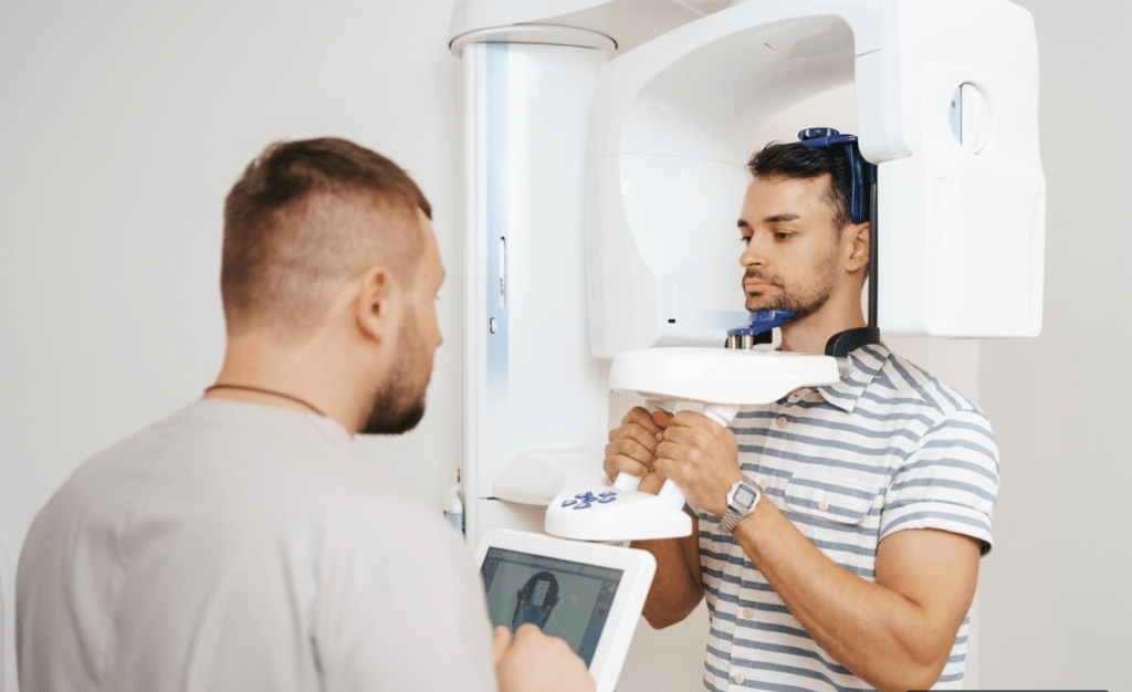
1. Panoramic X-rays
A panoramic X-ray is a wide-angle image that shows the entire mouth—teeth, upper and lower jaws, sinuses, and jaw joints—in one image. This X-ray type is commonly used in orthodontics to get a broad view of the patient’s dental structures, helping orthodontists assess the positioning of the teeth and the alignment of the jaws.
2. Cephalometric X-rays
Cephalometric X-rays are lateral head X-rays that show the side profile of the skull and teeth. This X-ray provides a comprehensive view of the facial bones, helping orthodontists analyze how the teeth relate to the skull and how the jaws align. It’s particularly useful in planning treatments like braces or jaw surgery.
3. Intraoral X-rays
Intraoral X-rays are taken inside the mouth to get detailed images of individual teeth. These X-rays are typically used to detect issues like cavities, tooth decay, and bone loss around the teeth, which are important considerations during orthodontic treatment.
4. Cone Beam CT (CBCT)
For more detailed 3D images, some orthodontists use cone beam CT scans. These provide a three-dimensional view of the teeth, bone, and soft tissues, making it easier to diagnose complex issues and plan intricate treatments, including implants or surgeries.
Each of these X-rays provides unique insights that contribute to a comprehensive treatment plan, ensuring that orthodontic care is as accurate and effective as possible.
The Role of X-Rays in Orthodontic Treatment Planning
Orthodontic treatment planning is an intricate process that involves understanding the structure of the teeth, jaw, and facial bones. Dental radiology services enable orthodontists to gather crucial information that cannot be assessed during a visual examination alone. Here’s how X-rays help in the process:
1. Assessing Tooth and Jaw Alignment
The most important aspect of orthodontic treatment is determining how the teeth and jaws are aligned. Dental radiology services allow orthodontists to see if teeth are positioned properly and whether there are any misalignments or malocclusions (bad bites). With the help of X-rays, orthodontists can plan exactly how much movement each tooth will need and decide on the type of braces or aligners that will be most effective.
2. Identifying Impacted Teeth
Sometimes, teeth don’t emerge from the gums as they should. Impacted teeth—teeth that are trapped beneath the gum line—can lead to misalignment and crowding. X-rays help orthodontists locate impacted teeth early in the treatment process, so they can plan for their proper movement or removal if necessary.
3. Monitoring Bone Development
The bones that support your teeth play a key role in orthodontics. X-rays allow orthodontists to monitor bone development and assess whether the jawbone is healthy enough to support teeth movement. This is especially important in younger patients whose bones are still developing. It also helps determine the best timing for initiating orthodontic treatment, as certain stages of bone growth may be more optimal than others.
4. Planning Surgical Interventions
In cases where traditional braces or aligners may not be sufficient, surgical procedures may be required to reposition the jaw. Dental radiology services such as CBCT scans give a precise 3D view of the patient’s skull and facial bones, allowing orthodontists to carefully plan the surgery and predict the outcomes more accurately.
5. Tracking Treatment Progress
X-rays aren’t just used at the beginning of treatment—they are also useful throughout the orthodontic process. Regular X-rays can help orthodontists track how well the teeth are moving and if the treatment plan is working as expected. If necessary, X-rays allow for adjustments to be made to the treatment to ensure the best results.
6. Evaluating Root Health and Bone Density
As teeth are moved during orthodontic treatment, it’s essential to ensure that the roots remain healthy and the bone density is sufficient to support the shifting teeth. X-rays help monitor root health, identify any potential bone loss, and ensure that the movement is occurring safely.
The Benefits of Dental Radiology in Orthodontics
Dental radiology services offer numerous benefits that enhance the efficiency and effectiveness of orthodontic treatments:
-
Early Detection: X-rays can reveal issues that might not be noticeable during a regular dental exam, allowing for early intervention.
-
Personalized Treatment Plans: By providing a comprehensive view of the mouth and jaws, X-rays enable orthodontists to create customized treatment plans based on the patient’s unique dental structure.
-
Faster and More Effective Treatment: With precise planning based on X-ray images, orthodontic treatment can be more efficient, leading to faster results with fewer complications.
-
Safety: The radiation used in dental X-rays is minimal and carefully controlled, making them safe for patients.
Conclusion
Dental radiology services are indispensable tools in modern orthodontics. X-rays provide orthodontists with a comprehensive view of a patient’s teeth, jaw, and bone structure, enabling accurate diagnoses and highly effective treatment plans. By using these imaging techniques, orthodontists can ensure that treatments are customized to the individual needs of each patient, ultimately improving the overall outcome.
If you are considering orthodontic treatment, rest assured that dental radiology services will play an integral role in guiding the entire process from start to finish, ensuring that your smile will be aligned in the safest and most efficient way possible.